What is kidney cancer?
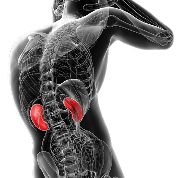
The kidneys are bean-shaped organs, and most people are born with two. Each kidney is about the size of your fist. The kidneys sit on either side of your spine. Their vital job is cleaning your blood by filtering waste products and balancing your body’s fluids.
Sometimes, abnormal cells grow in one or both kidneys. This abnormal cell growth is called kidney cancer. Here’s another way to put it: Kidney cancer develops when cells in the kidneys begin to grow and divide uncontrollably instead of in an organized manner, forming masses called tumors.
Kidney cancer is a serious health issue that affects thousands of people each year.
What causes kidney cancer?
The abnormal cell growth of kidney cancer typically occurs due to changes (called mutations) in genes that control cell growth and division.
There are numerous causes for these mutations. One is genetics. In other words, you inherited this predisposition from your parents. Other reasons are external factors, like smoking, alcoholism, and exposure to certain chemicals.
Patients are often referred to us following the incidental discovery of a kidney tumor, which may or may not be malignant (cancer).
What are the different types of kidney (renal) cancer
There are different types of kidney cancer. The most common ones are:
Renal cell carcinoma
Renal cell carcinoma, or RCC, is the most common type of kidney cancer, making up about 90% of all cases. The most frequent subtype of renal cell carcinoma is clear cell renal cell carcinoma or ccRCC.
Transitional cell carcinomas
These cancers affect the lining of the kidneys, the urine-collecting system (ureter), and sometimes the bladder. They are treated to prevent recurrence by removing the kidney, ureter, and part of the urinary bladder.
Patients with transitional cell carcinoma of the kidney require lifelong monitoring of the bladder, which is performed through a procedure called cystoscopy, as well as CT scans of the remaining kidney.
Wilm’s tumor
Wilm’s tumor is the most common type of renal cancer in children. However, it is quite rare. There are only 1 in 10,000 pediatric cases in the US. Wilm’s tumor can occur in one or both kidneys.
Types of benign (non-cancerous) kidney tumors
Not all kidney growths are cancerous. Some are benign, meaning they won’t spread to other parts of the body and are usually not life-threatening. Some types of benign kidney tumors are:
Renal adenomas
These are small, slow-growing, and non-cancerous.
Oncocytomas
Oncocytomas are uncommon, benign kidney tumors that can grow to a considerable size.
Angiomyolipoma
Angiomyolipoma is the most common type of benign kidney tumor, usually seen in women.
What are the risk factors for kidney cancer?
While the exact causes of kidney cancer aren’t always clear, several risk factors are known to increase your chances of developing this disease. Some of these risk factors are:
Smoking
Smoking is a major risk factor for renal cell carcinoma.
Alcohol
While the connection is not as strong as with smoking, alcohol consumption may play a role in increasing the risk.
Genetics
A family history of kidney cancer can increase your risk, suggesting a genetic link.
High blood pressure
Poorly controlled high blood pressure can damage the kidneys and increase the risk of kidney cancer.
Obesity
People with excessive weight are at increased risk of several cancers, including kidney cancer.
Exposure to certain chemicals
Long-term exposure to workplace chemicals like trichloroethylene or cadmium can increase the risk of renal cell carcinoma.
Being a man
Males are at a higher risk of developing kidney cancer than females.
Chronic kidney disease
People who have long-term kidney problems are at a higher risk of developing renal cell carcinoma.
What are the symptoms and signs of kidney cancer?
One of the tricky things about kidney cancer is that it often doesn’t show any signs or symptoms in its early stages. This is why it is helpful to distinguish between general symptoms and more specific warning signs.
Kidney cancer symptoms
General symptoms that might indicate a problem, though not necessarily kidney cancer, include:
- Feeling constantly tired (fatigue)
- Losing weight without trying
- A fever that keeps coming and going
Signs of kidney cancer
These are more specific signs that should prompt you to see a cancer specialist (oncologist) as they could be related to kidney cancer:
- Blood in your urine (hematuria)
- Ongoing pain in your lower back on one side
- A lump or mass you can feel in your abdomen or side
How is kidney cancer diagnosed?
Getting a kidney cancer diagnosis early gives you the best chance for successful treatment. Our urologists use several methods to confirm the diagnosis and see how far the cancer has spread. These include:
Urine and blood tests
Kidney cancer doesn’t always show up in blood work or urine tests. However, these tests can provide insights into your overall health and help your doctor rule out other potential health issues.
Imaging tests
Interventional radiologists use advanced imaging techniques to help urologists (kidney specialists) and oncologists (cancer experts) thoroughly examine your kidneys and identify potential tumors.
These interventional radiology tests include:
CT scans and MRIs: These scans create detailed pictures of kidney tumors.
We usually use intravenous dye to obtain high-quality CT scan images. The injected dye creates clear images needed to assess the extent of the mass, lymph node involvement, or if the tumor has invaded the blood vessel draining the kidney (tumor thrombus). This helps us decide on the appropriate treatment.
Ultrasounds: Ultrasounds use sound waves to create images to help find unusual growths on or in the kidneys.
Intravenous pyelograms: An intravenous pyelogram (IVP) is a specialized X-ray examination. Dye is injected into your veins to create clear images of your kidneys, ureters, and bladder, helping doctors diagnose urinary tract problems. However, IVPs have largely been replaced by newer imaging methods such as CT scans, MRIs, and ultrasounds.
Biopsy
This involves using a special needle to collect samples from the kidney mass and examine them for abnormal changes. Pathologists review the samples to confirm or reject the cancer diagnosis.
Although it sounds complicated, a needle biopsy is a relatively simple procedure. It is not even considered minimally invasive surgery.
In many cases, imaging alone is enough to guide treatment plans.
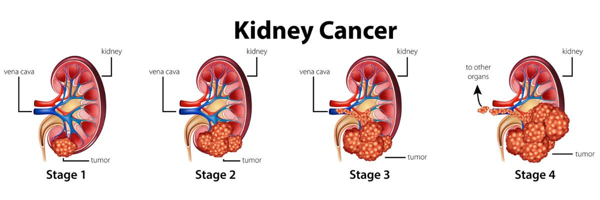
How do we rate how bad the cancer is?

Your healthcare team will use a standardized system to describe the stage (or level) of your kidney cancer. This staging system describes both the size of the tumor and its extent of spread from its original location in the kidney.
- Stage I: The tumor is small (7 cm / 3 in or less) and is only in the kidney.
- Stage II: The tumor is larger than 7 cm / 3 in but is still only in the kidney.
- Stage III: The tumor has spread to major blood vessels, surrounding tissue, or nearby lymph nodes.
- Stage IV: The tumor has spread beyond the kidney to the adrenal gland, distant lymph nodes, or other organs.
Is kidney cancer curable?
If kidney cancer is caught early, it’s often highly treatable. The 5-year survival rate for kidney cancer that hasn’t spread is excellent, usually over 90%.
Kidney cancer treatment options
The type of kidney cancer or cell type determines the best treatment plan. Generally, the success of the treatment depends on whether the cancer cells can be completely removed through surgery.
Surgery
Surgery is often the primary treatment option for kidney cancer. Fortunately, because each kidney functions independently, it’s often possible to remove one kidney while preserving the function of the other. The main surgical options are:
- Partial nephrectomy: This surgery removes only the tumor, preserving as much of the kidney as possible. The goal is to preserve as much of the kidney as possible so it can continue to function.
- Radical nephrectomy: This nephrectomy surgery removes the entire kidney. It’s usually done if the cancer is very large or has spread.
Targeted therapy
These medicines block the signals that help cancer cells spread, such as cutting off blood supply from their blood vessels or blocking specific proteins on cancer cells that help them grow and divide.
Immunotherapy
This treatment helps your body’s immune system find and attack cancer cells. Many medical oncologists (cancer experts) specialize in immunotherapy treatments for kidney cancer.
Ablation
Minimally invasive procedures, like radiofrequency ablation, which use heat to destroy cancer cells. Ablation does not destroy healthy tissue.
Active surveillance
We may recommend active surveillance when the tumor is small and growing slowly, especially in older patients. This means we closely monitor the patient and start treatment only when there is evidence that the cancer is growing. If the cancer has not spread beyond the kidneys, surgery to remove the tumor and part or all of the kidney may be the only necessary treatment.
Clinical trials and new treatments
Clinical trials are ongoing to test new treatments for kidney cancer. You should consult with your cancer specialist or visit a cancer center to explore the latest treatment options.
Finding the best kidney cancer care
For the best patient care, Tower Urology has assembled a strong care team, including urologists, radiologists, and cancer specialists, to help create a personalized treatment plan for each of our patients.
Why trust Tower Urology for your renal cancer care?
Tower Urology’s board-certified urological team has been a leader in effectively treating cancer for over two decades, with specialists trained in all aspects of prostate health.
Tower Urology is a proud affiliate of Cedars-Sinai Medical Center, ranked #1 in California and #2 nationwide by U.S. News & World Report. This partnership reflects our dedication to delivering the highest standard of urologic care alongside the best urologists in Los Angeles.
Our years of experience and access to Cedars-Sinai’s world-class facilities ensure that our exceptional and innovative urological care positions Tower Urology as a leader in Southern California.
We invite you to establish care with Tower Urology.
Tower Urology is conveniently located for patients throughout Southern California and the Los Angeles area, including Beverly Hills, Santa Monica, West Los Angeles, West Hollywood, Culver City, Hollywood, Venice, Marina del Rey, and Downtown Los Angeles.
Our services include treatment for bladder and urologic cancer, kidney cancer, prostate cancer, testicular cancer, and cancer fertility management.
Kidney Cancer FAQs

This is a renal cell cancer doctor with advanced training and expertise in diagnosing and treating kidney cancer. Our team includes urologic oncology and radiation therapy experts who specialize in genitourinary cancers (radiation therapy oncologists).
If you suspect kidney cancer, see your primary healthcare provider first. They will then refer you to one of our urologists or an oncologist specializing in kidney cancer for further evaluation and treatment. You may request a second opinion to confirm the diagnosis.
Kidney cancer prognosis varies greatly depending on the stage, type, and overall health of the individual. Early detection significantly improves the 5-year survival rate, which can be over 90% for localized kidney cancer. A medical oncologist can provide a more personalized prognosis.
Sources
Kidney Cancer Stages | Renal Cell Carcinoma Staging | American Cancer Society
https://www.cancer.org/cancer/kidney-cancer/detection-diagnosis-staging/staging.html
Targeted Therapy for Kidney Cancer │Targeted Therapy for Renal Cell Carcinoma | American Cancer Society
https://www.cancer.org/cancer/types/kidney-cancer/treating/targeted-therapy.html
Wilms Tumor and Other Childhood Kidney Tumors Treatment
https://www.cancer.gov/types/kidney/patient/wilms-treatment-pdq
Transitional Cell Cancer (Kidney/Ureter) Treatment
https://www.cancer.gov/types/kidney/patient/transitional-cell-treatment-pdq
Renal Cell Cancer Treatment
https://www.cancer.gov/types/kidney/patient/kidney-treatment-pdq