What is UPJ obstruction?
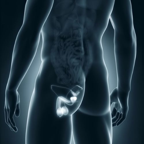
The ureteropelvic junction (UPJ) is the point at which the tube that carries urine from the kidney (the ureter) connects to the kidney. UPJ obstruction is a condition where the urine flow from the kidney is blocked at the point where these two structures meet.
This blockage can lead to urine buildup in the kidneys, causing swelling and potentially damaging the kidneys if left untreated. The condition can be congenital (present from birth) or may develop later in life.
Symptoms of UPJ obstruction can include flank pain, especially after consuming large amounts of fluid, urinary tract infections (UTIs), unexplained abdominal mass or swelling, kidney stones, and bloody urine.
While some cases may be mild and only require monitoring, more severe cases often necessitate surgical intervention through pyeloplasty. This procedure involves reconstruction of the blocked area to correct the abnormalities and restore normal urine flow.
This condition is one of the most common causes of hydronephrosis (kidney swelling) in newborns, but non-congenital UPJ obstruction can affect older children and individuals of any age.
What causes UPJ obstruction?
The diagnosis of ureteropelvic junction obstruction, or UPJ obstruction, indicates a blockage of urinary flow from the kidney down into the ureter. While most cases tend to be congenital, i.e., the person is born with it, symptoms tend not to manifest as anything clinically significant until much later in life.
Congenital blockage is usually related to three different conditions
- The ureter relies on the normal peristaltic (wave-like) movement of the muscles to propel urine along the ureteral tube. When the ureter develops an abnormal aperistaltic segment, the muscle bundles are ineffective in pushing the urine from the renal pelvis (the area at the center of the kidney where urine is collected) into the ureter.
- A high ureter insertion point into the renal pelvis can cause ineffective urine drainage from the kidney’s dependent portion.
- Crossing blood vessels occur when branch arteries from the main renal artery and vein cross behind the course of the UPJ, causing an intrinsic lesion within it.
Non-congenital causes include:
- Acquired conditions as a result of urine flowing backward from the bladder (known as vesicoureteral reflux)
- Scar tissue, often from previous surgeries, infections, or kidney stones
- Kinking or compression of the ureter by a blood vessel or other structure
- Benign polyps
- Cancers within the urinary tract
- Extrinsic compression, where cancers, growths, or pressure from other structures cause a blockage within the UPJ
How do we diagnose UPJ obstruction?
Most adult patients present with flank pain, which is exacerbated by increased fluid intake, caffeine, or medication such as diuretics. A CT scan with contrast can provide detailed information about the anatomy and function of the affected kidney. Modern imaging technology can also assess the presence of any crossing vessels.
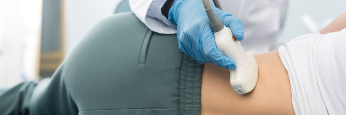
Ultrasound is a valuable diagnostic tool that enables the simultaneous detection of kidney stones. Furthermore, ultrasound is recommended to differentiate UPJ obstruction from other diagnoses in children and babies, or even via a prenatal ultrasound, thereby avoiding unnecessary radiation exposure from imaging studies such as X-rays.
After an initial CT or ultrasound exam, a nuclear renal scan with diuretic washout (using Lasix) can provide objective information about the degree of obstruction by evaluating the split kidney function between the two kidneys and the duration required for the kidneys to eliminate the injected isotope.
The T½ is the time it takes for the compound or isotope to be washed out of the kidney. A T½ of greater than 15-20 minutes usually implies a significant enough obstruction that requires further intervention.
When imaging and/or functional studies are unclear, a cystoscopy and retrograde pyelogram (involving the injection of contrast from the bladder into the kidneys) can confirm the diagnosis and identify the precise area of the blockage, allowing for the planning of a definitive repair.
Treatment is indicated when the patient experiences moderate to severe symptoms, damage to kidney function, recurrent stones or infections, or high blood pressure resulting from the blockage.
Treatment options for UPJ obstruction
Port placement for robotic pyeloplasty
A temporary measure to bypass the blockage is to place a plastic tube (double-J stent) with coils at the ends to prevent it from dislodging from the kidney. However, this is not a practical approach for addressing the blockage in most patients, as the stent needs to be replaced every 4 to 6 months.
Minimally invasive endoscopic techniques have proven effective in select individuals, depending on their underlying conditions and overall health status. Some of the benefits of endoscopic repair include less post-operative recovery time, avoiding incisions, and the option to have the procedure performed as an outpatient.
Endoscopic repair (endopyelotomy)
This procedure involves a full-thickness incision of the ureter at the site of the blockage. A ureteroscopy (an endoscopic procedure that looks directly into the ureter) is usually required to identify the area of blockage and make the incision using laser energy or balloon dilation with a cautery incision. The incision is carried from the lumen of the ureter out to the surrounding fat.
The opening subsequently heals over a special stent left in place for about 4-6 weeks. Endoscopic repairs are usually effective in the long term in only 60-70% of patients. In addition, almost half of the patients who undergo endoscopic repair may continue to have some degree of chronic flank pain after this procedure.
Dismembered pyeloplasty
The gold standard treatment for UPJ obstruction is a dismembered pyeloplasty, a technique in which the affected area is removed, and the renal pelvis is reconnected to the healthy, normal ureter.
This reconstruction was historically performed through an open surgery incision, usually through the flank. Laparoscopic pyeloplasty was developed as a minimally invasive alternative to the open technique. Laparoscopic pyeloplasty is associated with a shorter hospital stay, reduced pain medication, faster recovery, and better cosmetic results, while maintaining a 90% success rate comparable to that of formal reconstruction.
Similar to managing other urologic diseases, pure laparoscopic repair is technically challenging and has been surpassed by robotic assistance. Robotic pyeloplasty enables increased movement, dexterity, and precision that cannot be achieved with pure laparoscopic techniques.
Our minimally invasive surgical procedures are performed by surgeons who are fellowship-trained and recognized experts in the use of robotic technology to carry out complex reconstructions even after failed endopyelotomy and open repairs.
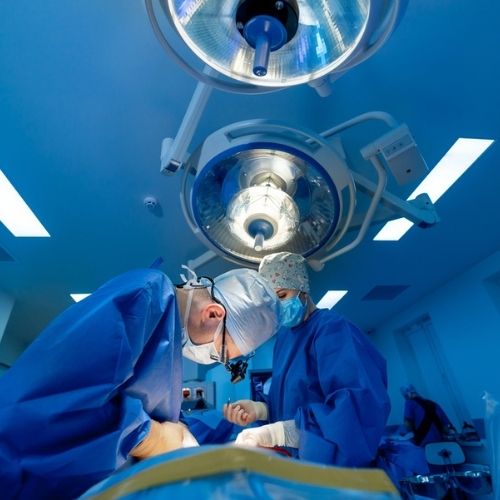
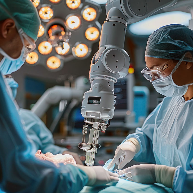
How is a dismembered pyeloplasty performed?
A dismembered pyeloplasty is now performed robotically (robotic pyeloplasty) under general anesthesia, typically takes around two to three hours, and requires a one-night stay in the hospital. The operation involves using robotic instruments placed in the abdomen through just three to four small incisions, each less than half an inch in size.
Why trust Tower Urology for the treatment of UPJ obstruction in Los Angeles?
Tower Urology’s board-certified urological team has been a leader in successfully treating UPJ obstruction for decades, with specialists trained in all aspects of urological care.
Tower Urology’s advantage lies in our unwavering commitment to providing world-class urologic care through advanced technology, personalized treatment plans, and a patient-centered approach. With a reputation for excellence and innovation, we deliver superior outcomes that set us apart as a leader in urologic health.
Tower Urology is a proud affiliate of Cedars-Sinai Medical Center, ranked #1 in California and #2 in the nation by U.S. News & World Report. This partnership reflects our commitment to delivering the highest standard of urologic care and related specialties with the best urologists in Los Angeles. Our years of experience and access to Cedars-Sinai’s world-class facilities and our exceptional, innovative, and state-of-the-art urological care make Tower Urology a leader in Southern California.
We invite you to establish care with Tower Urology.
Tower Urology is conveniently located for patients throughout Southern California and the Los Angeles area, including Beverly Hills, Santa Monica, West Los Angeles, West Hollywood, Culver City, Hollywood, Venice, Marina del Rey, and Downtown Los Angeles.
Services include treatment for enlarged prostate (BPH), erectile dysfunction (ED), low testosterone, male infertility, minimally invasive robotic surgery, mixed incontinence, overactive bladder (OAB), overflow incontinence, and kidney stones. We specialize in minimally invasive surgical treatment for issues relating to the urinary tract, including the kidney, bladder, and ureters.
Sources
Minimally invasive surgical options for ureteropelvic junction obstruction: A significant step in the right direction
https://pubmed.ncbi.nlm.nih.gov/19468425/
Adult Ureteropelvic Junction Obstruction: Insights with Three-dimensional Multi-Detector Row CT
https://pubs.rsna.org/doi/10.1148/rg.251045510