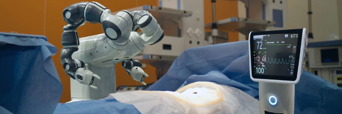
Posterior dissection of the seminal vesicles and the prostate
The seminal vesicles are cystic structures attached to the prostate and function to store and add nutrients to semen. The seminal vesicles are always removed with the prostate during surgery. A posterior approach enables energy-free and traction-free dissection of the nerve bundles that run along the sides and tips of the seminal vesicles.
The posterior approach also allows for perfect visualization of large or asymmetric seminal vesicles, situations which could be very complicated in an anterior approach. Additionally, this approach will enable us to bypass a large median lobe (a protrusion of the prostate into the bladder) without compromising visualization.
Developing the space of Retzius and anterior prostate dissection with sparing of the Endopelvic Fascia
This step drops the bladder and the prostate from the abdominal wall. It allows the surgeon direct visualization of the anterior prostate and the dorsal vascular complex. By sparing the Endopelvic fascia, an Intra-fascial dissection is more likely to be successful. See neurovascular bundle dissection below for more details.
Division of the prostate from the bladder by the bladder-neck sparing technique to preserve the internal sphincter mechanism
Two layers of bladder muscle fuse together the bladder and prostate in addition to fatty tissue. This area includes the internal urinary sphincter, which is composed of muscle fibers that play a role in the subconscious control of urinary function. In most patients, preservation of the bladder neck sphincter mechanism can result in early urinary continence. Proper preservation can also reduce the risk of strictures or scars at the bladder neck, commonly referred to as bladder neck contractures.
When the bladder neck is not spared, the subsequent large opening may necessitate a time-consuming reconstruction to narrow the opening. Larger openings also require a longer suture line and, therefore, may be more susceptible to leakage of urine from the anastomosis. Given all these factors and assuming there are no biopsy features putting the patient at risk for bladder neck involvement or the presence of a large median lobe, we always strive to preserve the bladder neck.
Dissection of the lateral prostatic fascia and sparing of the neurovascular bundles (NVB)
The nerve bundles carry neural information and blood flow into the deep pelvis. These structures are critical for both erections and urinary control after surgery. Multiple studies to date have shown a direct relationship between the degree of nerve sparing and post-operative potency and urinary control.
Contrary to what most patients believe, nerve sparing is not an all-or-nothing concept. Depending on the extent of the cancer, the nerve dissection can be individually tailored to the patient and their cancer.
Many factors come into the decision but include the risk of extracapsular extension based on pre-operative nomograms/risk-tables (link to separate section/post), the results of pre-operative T3 MRI of the prostate (link so section), the findings of a rectal exam under anesthesia, intra-operative findings and the ease of “peeling” the bundles away from the prostate.
The degree of precision and dexterity that is needed to dissect the nerve bundles away from the prostate is similar to what is required to “peel” the skin off of a tabletop grape (link to video).
Our goal is to remove all prostate and cancerous tissue clearly, but as important as a goal is to leave all non-prostate tissue as it was before the surgery. Many times, surgeons use the terms extrafascial, interfascial, and intrafascial to describe different techniques to dissect the prostate and the nerve bundles.
Extrafascial: This is also known as non-nerve sparing or wide-excision dissection. When there is either high suspicion that the tumor has penetrated the capsule wall deep into the extracapsular fatty tissue or is involving the nerve bundle itself, this may require us to resect the nerve bundle altogether with the prostate.
Typically, a preoperative MRI can determine the extent of involvement. In this type of dissection, the endopelvic fascia is incised deep near the levator ani muscles to carry out this type of dissection. In patients who are not interested in nerve preservation due to baseline erectile dysfunction, a wide excision may not be critical.
However, even in patients with clear evidence of extracapsular involvement, we may be able to do a graded dissection and spare some of the neurovascular bundle on the side affected with cancer in hopes of maximizing post-surgical potency. We use intra-operative frozen sections whereby we send tissue at the edge of the dissection to get a preliminary assessment of any tumor at the margin and subsequently guide our dissection based on those results.
We are also currently working on a novel technology that will enable the real-time identification of prostate tumors using fluorescent dyes targeting the cancer and near-infrared imaging, similar to what we have pioneered with kidney cancer surgery (link to Firefly). Although we are currently evaluating this technology in an animal model, we hope to apply this to clinical use soon and recruit patients into a clinical trial.
Interfascial dissection: The endopelvic fascia is incised, and the neurovascular bundles are spared posterolaterally to take some of the tissue around the prostate (periprostatic fascia) with the specimen. The component of nerves and vessels that can sometimes be found on the anterior aspect of the prostate are therefore not spared. As noted above, the clinical extent of the tumor has an impact of whether or not we decide to do this type of graded dissection.
Intrafascial dissection: This is the most delicate and precise type of dissection. Think of this as a custom-made, very “fitted” type of dissection. In this type of dissection, the endoplevic fascia, the neurovascular bundles, the periprostatic fascia, and Denonvillier’s fascia are all spared and left intact. The plane of dissection is directly guided on top of the prostate capsule. By preserving all these structures, we maximize the patient’s chance of regaining erectile function and urinary control after surgery.
Control of dorsal vascular complex (DVC)
As its name implies, the DVC contains an array of both veins and arteries that carry and drain blood from the penis. Due to the large amount of blood flowing in this structure, inadvertent injury can result in a significant amount of bleeding. The higher amount of blood loss associated historically with open prostatectomy techniques was related directly to this point of the procedure.
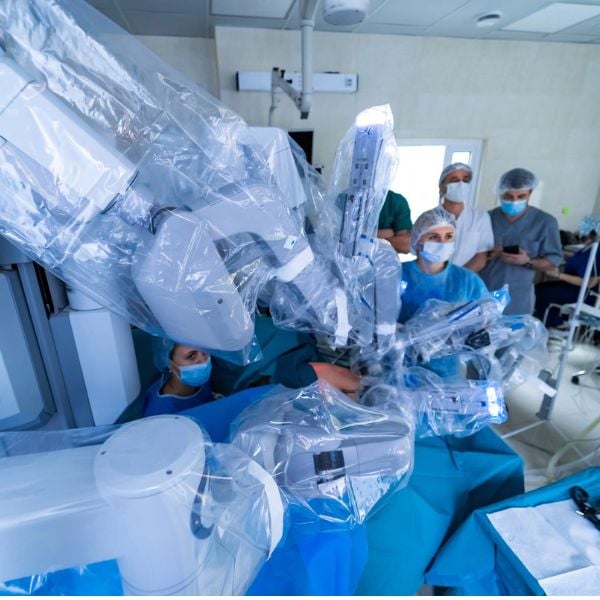
Fortunately, with the utilization of CO2 gas for laparoscopic and robotic surgery, venous bleeding from the DVC is no longer a major issue, as the gas can provide passive pressure and prevent oozing from this area. Two methods are used to control the DVC: either suture ligation or endoscopic stapling.
One of our preferred methods of control is to use an endoscopic stapler. As we have previously published, endoscopic stapling allows for a consistent, efficient, and reliable method for getting control without risking a positive margin at the apex of the prostate.
Preparation of Apical Urethra – preserve length and muscle fibers
Prostates come in all sizes and shapes. Some are smaller, some are bigger. Most of the variation in shape is usually seen at the apex. The apical prostate surrounds the urethra near the external sphincter complex. The goal at this point of the operation is to achieve a long and thick urethral stump that will subsequently be reconnected to the bladder neck.
There is a fine line between dissecting too deeply (the levator muscles and the external sphincter complex can be inadvertently damaged) and too shallowly (risk leaving prostate tissue behind).
Posterior Reconstruction
Also known as a Male sling, Rocco reconstruction, or Rhabdosphincter reapproximation. This technique provides posterior support for the sphincter complex, similar to a hammock, and prevents the urethra from slipping further down into the pelvis during activity, which may lead to leakage, such as during coughing, sneezing, or laughing.
The reconstruction also brings the bladder down into a supported position and therefore removes any tension on the completed urethra-bladder anastomosis. Most experts agree that this type of reconstruction leads to shorter recovery times for urinary control.
Urethrovesical anastomosis
The goal at this point of the procedure is to create a watertight and tension-free connection between the urethra and the prostate. One of the most significant advancements to our practice has been the ability to utilize a barbed suture (V-Loc, Covidien) that maintains the reapproximated ends without allowing any gaps between the tissue edges. As a result, for years now, we have avoided using any post-operative drains unless we are dealing with a very large bladder neck or a reconstructed bladder neck.
Pelvic Lymph Node dissection: Removal of pelvic lymph nodes – extended dissection for patients at greater risk of involvement based on biopsy features, clinical exam and PSA
Even though pre-operative CT or MRI may not show evidence of regional lymph node involvement, the accuracy of these imaging tests is only around 80%. The decision to identify and remove the lymph nodes that drain the prostate during prostatectomy is made on an individual basis. We calculate the risk of lymph node involvement based on widely available nomograms and risk calculators such as D’Amico criteria, Partin Tables, CAPRA score, or MSKCC calculator.
Low-risk patients (for example, those with PSA < 10, Gleason 6, or clinical T1c) do not benefit from a lymph node dissection. Intermediate-risk patients and High-risk patients, on the other hand, may benefit from a lymphadenectomy. It is believed that an extended lymph node dissection (in both lymph node negative and positive patients) may lead to the removal of undetected micrometastases, and therefore improve the survival of patients undergoing prostatectomy.
There is a growing body of evidence suggesting that the greater the number of lymph nodes removed, the more beneficial. However, no uniform consensus exists regarding the limits of the boundary of the dissection or the minimum number of lymph nodes that should be removed. Historically, the detection of suspicious lymph nodes at the time of radical prostatectomy led many surgeons to abandon the operation with the belief that regional lymph node involvement was a sign of widespread metastatic disease and therefore associated with poor prognosis.
Patients would subsequently be referred for treatment with hormones and/or radiotherapy. However, there have been several recent studies showing very reasonable cancer-specific survival rates even in patients with lymph node-positive disease at the time of radical prostatectomy. A recent publication from the European Journal of Urology analyzed the Munich cancer registry and found that patients with lymph node-positive disease who did not have their operation aborted on average had a 20% improvement in survival compared to those who had their operation aborted.
A family member was struggling with testicular pain for years and had concerns about fertility. I am so grateful that we were referred to Dr. Houman. He is very thoughtful and knowledgeable and has great bedside manner. He diagnosed a varicocele and recommended microsurgical varicocelectomy and we are so happy with the results.
I highly recommend Dr. Houman.
Dave R.
★★★★★
Great doc you can trust. He's very up on all the new and modern modalities to help you and keep you healthy. Great choice and nice guy.
Ilan K.
★★★★★
Amazing urologist. Go to expert for men?s health. I refer patients to him as often as possible!
Marilyn B.
★★★★★
How did a woman with a lifetime of recurrent bladder infections wind up with a urologist whose specialty is male fertility?
It happened like this...Dr. Houman was recommended to my husband when he began having the prostate problems that can devastate older men. Dr. Houman solved his urinary difficulties, and I'm convinced, kept him from the fate of a constant catheter.
Naturally, I jumped at the chance to be his patient, and have been, for the first time in my life, free for several months of any infections.
But the best part of being Dr. Houman's patient is this. When you are in trouble, he will keep in constant touch with you...he does not isolate himself behind a wall of nurses. Naturally, I recommend him highly,
Marilyn B.
Afshin A.
★★★★★
There are doctors who go into medicine for the paycheck and then there are doctors like Dr. Justin Houman. From the very first appointment you can feel that he truly cares. He?s not just knowledgeable, he?s patient and genuinely passionate about what he does. You can tell he?s in this to help people not just to treat symptoms but to make a real difference in their lives. I felt seen, heard and supported every step of the way. Thank you Dr. Houman for going above and beyond for your patients. You are a rare kind of doctor and I?m so grateful I found you.
Paul S.
★★★★★
Dr. Houman is amazing and diagnosed my issue immediately. instead of a surgery I thought for certain I would need, he prescribed medication that solved the problem.
he was very detailed in his meetings with him and his follow up exam perfect whereby he asked if I had any other issues.
I highly recommend him to anyone as he cares and does a thorough assessment and treatment.
Gregg D.
★★★★★
Simply, Dr. Josephson gave me my life back. He is the best there is. Great bedside manner, cutting edge technology, great staff, and delivered on everything he told me would happen. Thank you Dr. Josephson!!
Aaron M.
★★★★★
Dr. Houman and his staff are incredibly capable. Provide customer service like no medical practice I have been to, very attentive. Addressed all my issues, spent time listening to me, and explained exactly what has been troubling me. Cant thank him enough for his care, kindness, and diligence
Christopher G.
★★★★★
The level of care and attention Dr. Houman gives during a appointment is second to none. He explained different levels of treatment, answered all my questions and never once did I feel time was an issue. Thank you Dr. Houman for making me feel I was a top priority during my appointment.
Jordan E.
★★★★★
So glad I came across Dr. Houman. He was extremely attentive in taking care of all my needs. He has an incredible attention to detail and took the time to listen to my particular needs. I definitely see him being my physician for life!
Ariel R.
★★★★★
So kind and considerate, WONDERFUL bedside manner! Did wonders for our sex life! Thank you for everything!!
Vishal G.
★★★★★
Dr. Houman is thorough and really knows his stuff.
Josh G.
★★★★★
Dr. Houman absolutely sets the standard when it comes to patient care and bed side manner. I made an appointment for a very concerning topic and was full of worry. Not only was the process of making an appointment with him easy, but the customer service was impeccable. His assistant was joyful and happy to help. The experience only got better from there. The moment I met Dr. Houman I was quickly put at ease. He patiently answered any of my questions and concerns as well as explain things in a way that made them easy to understand. I have worked closely with many physicians but he goes above and beyond. He truly has a gift and I highly recommend him for any of your urological needs. Can't thank him enough for his kindness and exceptional professionalism.
John M.
★★★★★
I've been working with Dr. Houman for a few months now after a complication from a surgery. I was referred to Dr. Houman and he assured me that we would get the issue resolved. I had a lot more going on than initially known. Each problem has been addressed and it seems I'm on my way to normal. Healing takes time. It's important to have a caring, professional and diligent Doctor and staff on your team. The nurses working with Dr. Houman are knowledgable and highly skilled. I recommend Dr. Houman and his staff.
Samuel G.
★★★★★
Dr. Houman is not only extremely professional and friendly, but highly knowledgeable in his field. I was unsuccessful after seeing two other physicians, but Dr. Houman knew the issue and addressed it within 1 month of meeting him!
New Patient Portal Instructions – Tower Urology Los Angeles
Tower Urology is excited to introduce you to our patient portal. On the portal, you can:
Securely communicate with your doctor
View your health information
And even manage your family’s care!
Sign up for an account to access the above and more anytime and anywhere! It’s an easy way to stay connected and communicate with Tower Urology about your healthcare.
To gain access to our new patient portal, please get in touch with Tower Urology at (310) 854-9898