What is a robotic pyeloplasty, and what does it treat?
A robotic pyeloplasty is a minimally invasive surgery that fixes a blockage where your kidney connects to the tube (called the ureter) that drains urine down to your bladder.
A ureteropelvic junction (UPJ) obstruction is a blockage at the point where the kidney drains into the ureter. It can cause symptoms including pain or infection, or the formation of a kidney stone.
The most effective way to treat a UPJ is with robotic pyeloplasty — minimally invasive surgery that clears the blockage and preserves kidney function.
At Tower Urology, we perform robotic pyeloplasty for patients throughout Southern California seeking advanced care with a gentle approach. In this article, we cover what a UPJ obstruction is, how we diagnose it, what happens during robotic pyeloplasty, and what recovery looks like.
What is a UPJ obstruction?
A UPJ obstruction is a blockage in the tube (called the ureter) that drains urine from the kidneys to the bladder. The kidneys are responsible for making urine, which collects in the renal pelvis, a funnel-shaped structure. From there, the urine flows into the ureter, a thin, muscular tube about 8 to 12 inches long that carries it to the bladder.
The point where the kidney meets the ureter is known as the ureteropelvic junction (UPJ), where a blockage can occur. Most people are born with a UPJ obstruction, but others develop it due to:
- Scar tissue from prior surgery
- Previous infections that narrowed the ureter
- A crossing blood vessel pressing on the ureter
- A kink or abnormal bend in the ureter
- Less commonly, a growth near the junction
Left untreated, a UPJ obstruction can cause kidney stones, recurring infections, chronic flank pain, and gradual loss of kidney function. Robotic pyeloplasty removes the blockage and restores a clear, healthy pathway for the urine to drain.
What are the symptoms of UPJ obstruction?
The most common symptoms of UPJ obstruction are persistent flank or abdominal pain, recurrent urinary tract infections (UTIs) or kidney infections, blood in the urine (hematuria), and vomiting.
But any of the following can indicate a UPJ obstruction.
Persistent flank pain
A dull, sharp, or crampy ache between the ribs and hip on one side, often worse after drinking fluids.
Blood in the urine (hematuria)
Visible pink or red urine, or blood detected on a lab test.
Recurrent UTIs or kidney infections
Repeated infections on the same side, often with fever, chills, or back pain.
Nausea and vomiting
Especially during a pain flare, when kidney pressure triggers a strong body response.
Kidney stones
Formed when urine sits in the kidney too long. When stones appear alongside a swollen kidney, it may indicate an UPJ obstruction.
Symptoms can come and go and can be easily mistaken for other problems. Additionally, some individuals have no symptoms at all, and the obstruction is found incidentally on imaging. Known as silent obstructions, they can still cause kidney damage over time, even without symptoms.
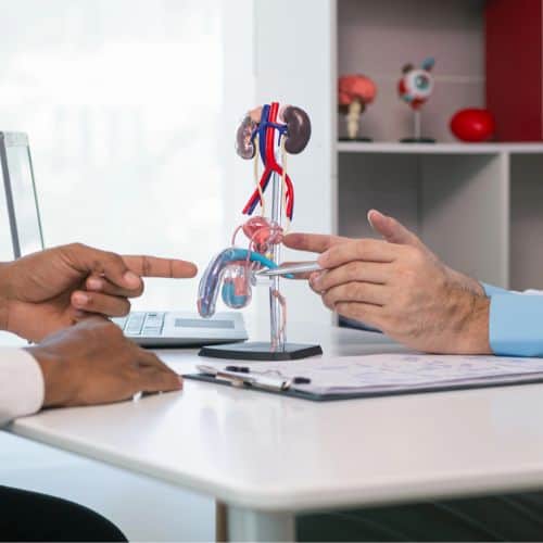
How is a UPJ obstruction diagnosed?
Diagnosis involves a combination of lab work and imaging. At Tower Urology, our urologists review your full medical history, prior surgeries, and medications before recommending specific tests, because these details shape both diagnosis and treatment, including whether a pyeloplasty procedure is appropriate for you.
Common diagnostic tests include:
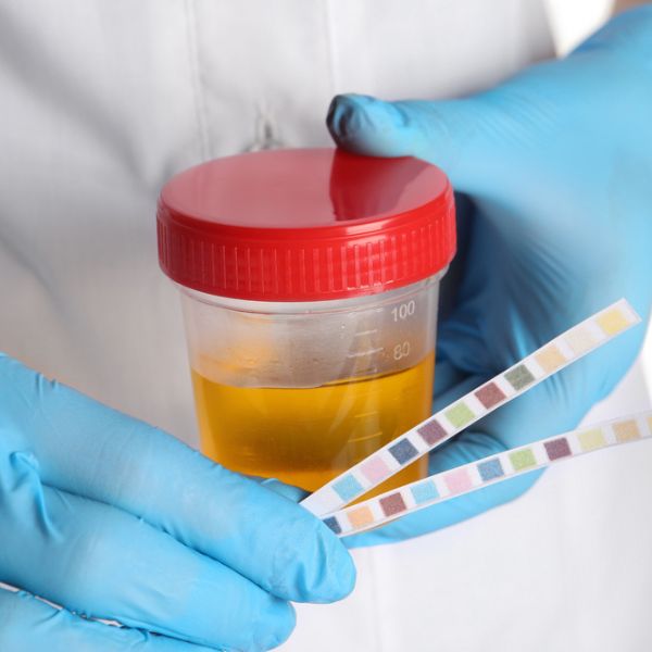
Blood and urine tests
Laboratory tests that check for infection, measure kidney function, and evaluate how well the kidneys are filtering waste.
Renal ultrasound
An imaging test that uses sound waves to create pictures of the kidneys, revealing any swelling or possible blockages.
CT urogram
A type of X-ray imaging that produces detailed cross-sectional pictures of the kidneys and ureters, pinpointing the exact location and severity of a blockage.
Cystoscopy
A procedure that uses a thin, flexible camera inserted through the urethra to directly view the inside of the bladder and ureter.
MAG-3 renal scan (diuretic renogram)
A nuclear medicine test that tracks a small amount of radioactive tracer to see how well each kidney drains and how much each contributes to overall kidney function.
MRI urogram
A radiation-free imaging test that uses magnetic fields to create detailed pictures of the kidneys and urinary tract, often used in younger patients.
These tests can not only show whether a blockage exists but also how serious it is and how much the affected kidney is still contributing to urination.
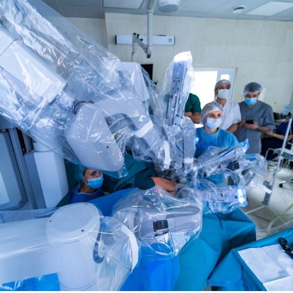
How is a robotic pyeloplasty performed?
Robotic pyeloplasty is the gold-standard surgical treatment for UPJ obstruction. Using a console in the operating room to operate the da Vinci robotic system, our surgeons make a few tiny incisions in the abdomen and work through those openings using robotic arms and a high-definition 3D camera.
Then our surgeon removes or repairs the narrowed or blocked section and reconnects the healthy parts so that urine can flow freely again. Because the incisions are so small, most people recover much faster and with less pain than after traditional open surgery.
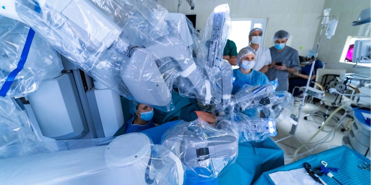
The da Vinci robot platform provides advantages over traditional open pyeloplasty procedures, including exceptional precision, magnified visualization, and instrument control that offers many benefits, such as:
- Smaller incisions, sutures, and significantly less scarring
- Less blood loss during the procedure
- Less post-operative pain
- Shorter hospital stay (typically one night)
- Faster return to normal activities
- Success rates consistently above 95%
During the procedure, the narrowed or scarred section of the ureter is removed, and the healthy, open end is directly reattached to the renal pelvis. This creates a wide, unobstructed connection that allows urine to drain normally.
A small internal stent is then placed temporarily to support healing and keep the tube open — this is typically removed 4-6 weeks later. Finally, the small laparoscopic incisions are closed, and the patient is moved to recovery.
For patients with a crossing blood vessel causing the obstruction, the robotic approach is especially valuable as it allows our surgeons to reposition the ureter away from the crossing vessels with the precision needed to avoid injury.
Is there a single-port robotic pyeloplasty option?
Yes! A single-port robotic pyeloplasty is a version of the same kidney-drainage repair, but instead of making several small incisions for the robotic instruments, the surgeon performs the entire procedure through a single small opening (often near the belly button). All the camera and surgical tools enter through that single port, then fan out inside the body to fix the blockage where the kidney meets the ureter.
The main advantage is that fewer incisions usually mean less scarring, often less pain, and a more cosmetic result, since there is essentially one hidden scar instead of several. Many patients also tend to recover and return to normal activities a bit more quickly.
Does a single-port robotic pyeloplasty require a more experienced urological surgeon?
Yes, it does. A single-port surgery is technically more demanding than the standard multi-port approach, so it tends to be performed by surgeons with the most advanced robotic experience. These include the fellowship-trained surgeon at Tower Urology.
For that reason, single-port pyeloplasty is most often performed at centers and by surgeons, such as the fellowship-trained urological surgeons at Tower Urology. They have a high volume of robotic cases under their belt and have been specifically trained on the single-port platform. If you are weighing options and are not a patient with Tower Urology, it is reasonable to ask a surgeon how many single-port procedures they have performed and what their outcomes have been.
What is robotic pyeloplasty recovery like?

After a robotic-assisted pyeloplasty, most patients spend one night in the hospital and go home the following day. A catheter is placed during surgery to drain urine and is often removed 1-2 days after surgery. Pain is manageable with oral medication, and many people feel well enough to move around the house within the first day or two.
A general recovery timeline looks like this:
1-2 weeks
Rest at home; light walking encouraged. Avoid heavy lifting (anything over 10 pounds).
3-4 weeks
Most patients return to desk work or light daily activity.
4-6 weeks
The internal ureteral stent is removed in a quick office procedure. Most patients resume full activity shortly afterward.
3 months
Imaging at a follow-up appointment can confirm the repair is open and draining properly.
As with any surgery, there are risks to be aware of, including bleeding, clots, infection, urine leak, or, rarely, re-narrowing of the repair site, and in very rare cases, the need for a blood transfusion.
Your surgeon will discuss these with you in detail before your procedure so you can make a fully informed decision.
The long-term outlook after robotic pyeloplasty is excellent. The vast majority of patients experience complete pain relief and a permanent resolution of the obstruction. When the surgery is performed before significant kidney damage has occurred, the affected kidney often recovers meaningful function.
Why Trust Tower Urology with your urinary health?
If you have been diagnosed with a UPJ obstruction or if you have experienced persistent flank pain, recurrent infections, or kidney swelling, our team at Tower Urology is here to help. We offer expert evaluation, precise urologic surgery, and personalized healthcare for patients across Southern California.
We invite you to establish a care plan with Tower Urology. Please request an appointment online or call us at (310) 854-9898.
Tower Urology is a proud affiliate of Cedars-Sinai Medical Center, ranked #1 in California and #2 nationwide by U.S. News & World Report. Our years of experience and access to world-class facilities ensure exceptional urological care.
Tower Urology is conveniently located for patients throughout Southern California and Los Angeles, including Beverly Hills, Santa Monica, West Los Angeles, West Hollywood, Culver City, Hollywood, Venice, Marina del Rey, Burbank, Glendale, and Downtown Los Angeles.
Frequently asked questions about robotic pyeloplasty
The procedure typically takes between two and three hours, depending on the complexity of the obstruction and whether a crossing blood vessel is involved.
Yes, a pyeloplasty with the da Vinci system is a robotic surgery that must be performed under general anesthesia, with the patient positioned carefully on the operating table. Our team reviews your health history in advance to ensure you are a safe and suitable candidate.
In most cases, yes. Robotic pyeloplasty is a medically necessary procedure when a UPJ obstruction is confirmed. Our team works with your insurance provider and walks you through what to expect regarding coverage and costs.
For mild obstructions with good kidney function, careful monitoring is sometimes appropriate. However, most confirmed UPJ obstructions benefit from surgical correction before kidney function declines, as this can lead to kidney failure and necessitate a nephrectomy (kidney removal).
Re-obstruction is uncommon after robotic pyeloplasty, occurring in fewer than 5% of cases. When it does happen, additional treatment options are available. Follow-up imaging at three months helps us confirm that the repair is working well.
Sources
Robotic-assisted pyeloplasty for ureteropelvic junction obstruction
https://csurgeries.com/video/robotic-assisted-pyeloplasty-for-ureteropelvic-junction-obstruction/
Advances and Trends in Pediatric Minimally Invasive Surgery
https://www.mdpi.com/2077-0383/9/12/3999
Future of robotic surgery
https://journals.lww.com/journalppo/abstract/2013/03000/future_of_robotic_surgery.2.aspx